Yesterday I attended the second dental symposium on surgical microscopy hosted by Zeiss. I think these symposia are great as they bring together some of the most skilled dental surgeons in the world to talk on the advances in micro-surgery. Its quite simple, the better you can see what you are doing the better you can do the surgery.
The first speaker was Prof. Krejci on the ‘Geneva Concept’. In summary decay and gum disease is a highly infectious incurable chronic disease and we acquire the bacteria responsible for it at birth. Therefore since we can’t cure it we need to focus on prevention and conservation of damaged tissues. To quote “There is no such thing as a permanent restoration, they are all temporary until the last one”. The easiest and most cost-effective treatment is early intervention and the use of adhesive materials such as resins and composites. With the use of microscopy we can preserve tooth tissue because it’s so much easier to see small defects and cavities in the teeth and precisely repair them.
Dr Rino Burkhardt’s subject was ‘Minimally invasive periodontal surgery and its effects on wound healing’. In essence success depends on maintaining the microscopic blood supply to promote rapid healing and was best achieved by adopting the skills from the other surgical disciplines such as eye and microvascular surgery.
Dr Domenico Massironi (one of my personal heroes) gave a beautiful presentation on the use of surgical microscopes in aesthetic dentistry. Only with improved vision can we truly assess the quality of our work and achieve the precision that our patients deserve. Check out his video (its only in Italian but that does not matter watch it anyway)
In the afternoon followed three excellent lectures by Prof. Gilberto Debelian, Dr Tony Struttman and Dr Tomas Lang on root fillings, retreating failed root fillings and micro-invasive dentistry. If a tooth can be reasonably saved there are huge advantages to the patient than losing it and hoping that an implant will perform any where as well.This may not have been so 15 years ago but microscopy now allows us to literally look down the root of a tooth which was impossible before then. It’s hard to out engineer nature.
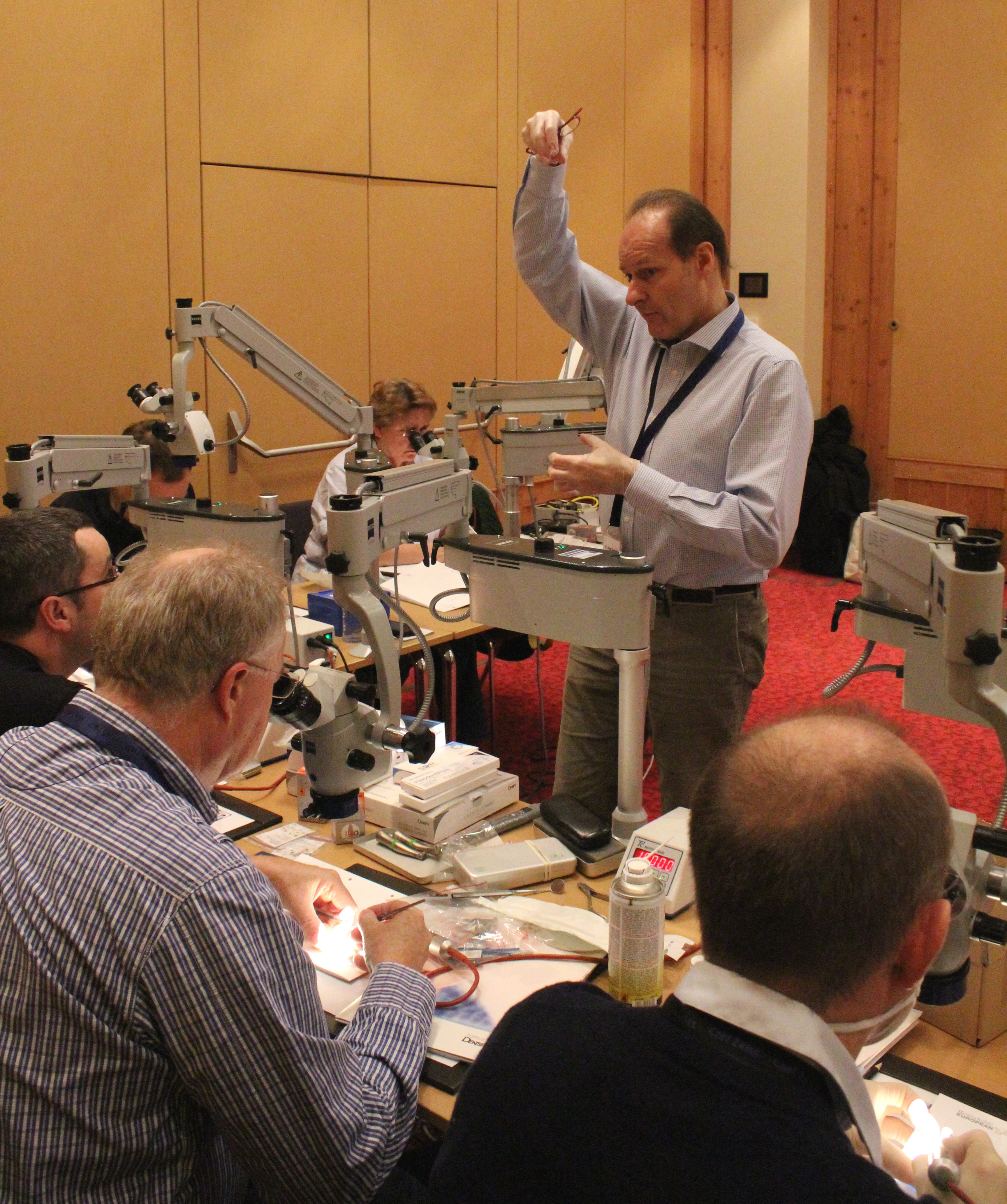
The final presentation was from Oscar von Stetten on using the microscope to document our treatment and communicate what we see down the microscope to colleagues and patients. If our patients truly understand what we as clinicians are trying to achieve the better the treatment outcomes. Below is a picture of a cracked back tooth which helped the patient understand why it hurt every time they bit on it.

